fredag den 26. juli 2019

Complex V is down in ME - does it also explain Electromagnetic Hypersensitivity?

Complex V

Complex V (also known as ATP synthase or F0F1ATPase) is working less efficiently in cells from ME patients (1).


Chemical Intolerance and Electromagnetic Hypersensitivity in ME

ME patients have chemical intolerance (CI), also knowns as multiple chemical sensitivity (MCS). 

I have asked the question:
Complex V is down in ME - does it explain Chemical Intolerance?
http://followmeindenmark.blogspot.com/2019/07/complex-v-is-down-in-me-does-it-explain.html

Several ME patients also have electromagnetic hypersensitivity (EHS). It has been hypothesized that EHS and CI are two etiopathogenic aspects of a unique pathological disorder (2, 3).

Maybe ME, CI and EHS are a unique "Complex V-disease"?


Electromagnetic Fields and Complex V

Humans are exposed to electromagnetic fields (EMFs) from: Radio and television stations and receivers, radar, computers, Wi-Fi antennas, mobile phones, microwave ovens, and many devices used in medicine and industry. Our blood is just under the skin and is exposed to EMFs all the time.

Lassaivia et al have investigated peripheral blood lympho-monocytes exposed to EMFs at 1.8 GHz frequency and 200 V/m electric field strength. Respirometric measurements of mitochondrial activity in intact lympho-monocytes showed:
  • An in increase of the resting oxygen consumption rate after 20 h of exposure, which was coupled to a significant increase of the FoF1-ATP synthase-related oxygen consumption.
  • At lower time-intervals of EMFs exposure (i.e. 5 and 12 h) a large increase of the proton leak-related respiration was observed which, however, recovered at control levels after 20 h exposure.
  • No significant variations in the mitochondrial mass/morphology was observed in EMFs-exposed lympho-monocytes.
  • Altered redox homeostasis was shown in EMFs-exposed lympho-monocytes, which progressed differently in nucleated cellular subsets.

The results suggest the occurrence of adaptive mechanisms put in action, likely via redox signaling, to compensate for early impairments of the oxidative phosphorylation system caused by exposure to EMFs (4).


ME and Complex V

Fisher et al have investigated lymphoblasts from ME patients and found specific mitochondrial respiratory defects and compensatory changes (5).

Respirometry of immortalized ME patient lymphocytes (lymphoblasts) showed:
  • Mitochondrial ATP synthesis by Complex V is inefficient, representing a significantly lower proportion of the basal mitochondrial respiratory activity.
  • Absolute ATP synthesis rates (pmol/min) were not significantly lower than in control cells, while glycolysis rates and steady state ATP levels were unchanged.
  • There was a significant increases in maximum respiratory capacity, including Complex I activity.
  • “Nonmitochondrial” O2 consumption by other cellular enzymes was also elevated. 

The results suggest that ME/CFS cells compensate for the reduced efficiency of ATP synthesis by upregulating mitochondrial respiratory capacity.

The mitochondrial membrane “mass” and genome copy number were unchanged. Thus ME/CFS cells do not have “more” mitochondria, but their mitochondria have greater respiratory capacity. This increased capacity is underutilized because of the Complex V defect, so that the respiratory “spare capacity” was increased and the mitochondrial membrane potential was elevated (5).

I have a new question:
Can continous exposure from electromagnetic fields in the environment put further strain on a compromised complex V in ME patient cells?


A magnetic field is able to influence Complex V

Interestingly, a team of Japanese scientists have succeeded in attaching magnetic beads to the stalks of F1 -ATPase isolated in vitro, which rotated in presence of a rotating magnetic field. F1 -ATPase synthesized ATP from ADP and Pi when rotated in a clockwise direction at a rate of about 5 molecules per second. Additionally, ATP was hydrolyzed when the stalks were rotated in the counterclockwise direction or when they were not rotated at all (6, ref 26 in ref 7).


References

1) Missailidis, D.; Annesley, S.J.; Allan, C.Y.; Sanislav, O.; Lidbury, B.A.; Lewis, D.P.; Fisher, P.R. An isolated Complex V defect and dysregulated mitochondrial function in immortalized lymphocytes from ME/CFS patients. Submitted 2019.

Specific Mitochondrial Respiratory Defects & Compensatory Changes in ME/CFS Patient Cells
https://www.youtube.com/watch?v=SjK39QCPeeY

2) Belpomme DCampagnac CIrigaray P. : Reliable disease biomarkers characterizing and identifying electrohypersensitivity and multiple chemical sensitivity as two etiopathogenic aspects of a unique pathological disorder. Rev Environ Health. 2015;30(4):251-71. doi: 10.1515/reveh-2015-0027. https://www.ncbi.nlm.nih.gov/pubmed/26613326

3) De Luca CThai JCRaskovic DCesareo ECaccamo DTrukhanov AKorkina L   Mediators Inflamm.: Metabolic and genetic screening of electromagnetic hypersensitive subjects as a feasible tool for diagnostics and intervention.  2014;2014:924184. doi: 10.1155/2014/924184. Epub 2014 Apr 9. https://www.ncbi.nlm.nih.gov/pubmed/24812443

4) Lassaivia et al: Exposure to 1.8 GHz electromagnetic fields affects morphology, DNA-related Raman spectra and mitochondrial functions in human lympho-monocytes. PLOS ONE. February 20, 2018.https://doi.org/10.1371/journal.pone.0192894

5) International research symposiym: Mitochondrial function and signalling “Specific mitochondrial respiratory defects and compensatory changes in immortalized ME/CFS patient lymphocytes.” Speaker: Professor Paul Fisher, Latrobe University, VIC, Australia
https://emerge.org.au/wp-content/uploads/2019/03/2019-Symposium-Programme.pdf

6) Itoh et al: Mechanically driven ATP synthesis by F1-ATPase. Nature. 2004 Jan 29;427(6973):465-8. https://www.nature.com/articles/nature02212

7) Neupane et al: ATP Synthase: Structure, Function and Inhibition. De Gruyter 2019.
https://www.degruyter.com/view/j/bmc.2019.10.issue-1/bmc-2019-0001/bmc-2019-0001.xml

tirsdag den 23. juli 2019

Complex V is down in ME - does it explain Chemical Intolerance?

Complex V

Complex V (also known as ATP synthase or F0F1ATPase) is working less efficiently in cells from ME patients (1).

The ATPase Inhibitory Factor 1 (IF1) is the physiological inhibitor of the mitochondrial ATP synthase (complex V). IF1 is a mitochondrial protein with very short half-life. It is tissue-specifically expressed and primarily controlled at posttranscriptional levels. Overexpressing of IF1 leads to inhibition of the ATP synthase and the reprograming of energy metabolism to an enhanced glycolysis. This reprogramming may protect cells from cytotoxic insults (2).

Figur 1B from reference 2 shows how IF1 binds the ATP synthase and inhibits ATP synthesis. Figur 1A: The gene ATP5IF1 encodes IF1. In mice IF1 is degraded by immediate early response gene X-1 (IEX-1). The human homolog af IEX-1 is immediate early response 3 (IER3), but IER3 do not degrade IF1 (2).
https://www.ncbi.nlm.nih.gov/pmc/articles/PMC6156145/figure/F1/


Chemical intolerance - is IF1 involved?

ME patients have chemical intolerance (CI), also knowns as multiple chemical sensitivity (MCS). Fx, ME patients do not tolerate smoke and exhaust fumes.

Polycyclic aromatic hydrocarbons (PAHs) are found in the environmental contaminants. It has been hypothesized  that dysregulation of the aryl hydrocarbon receptor is involved in chemical intolerance (3).

Benzo[a]pyrene (B[a]P) is a prototype molecule of polycyclic aromatic hydrocarbons. B[a]P induce IF1 expression and metabolic reprogramming towards glycolysis in rat liver cells. The process may also involve β2-adrenergic receptor and aryl hydrocarbon receptor activation (4).


An external file that holds a picture, illustration, etc.
Object name is 41598_2017_269_Fig6_HTML.jpg

Figure 6 from reference 4: A proposed model for the B[a]P-mediated metabolic reprogramming and its role in cell fate determination in F258 rat hepatic epithelial cells.
https://www.ncbi.nlm.nih.gov/pmc/articles/PMC5428028/figure/Fig6/

Can PAH-induced IF1 upregulation put further strain on a compromised complex V in ME patient cells?

Can auto-antibodies against adrenergic receptors upregulate IF1 activity in ME patient cells?

Mutant mice with an active IF1 are partially protected from cytotoxic insults - such as quinolinic acid (2). Interestingly quinolinic acid is a kynurenine metabolite which may be dysregulated in ME patients according to the IDO metabolic trap hypothesis (5).


References

1) Missailidis, D.; Annesley, S.J.; Allan, C.Y.; Sanislav, O.; Lidbury, B.A.; Lewis, D.P.; Fisher, P.R. An isolated Complex V defect and dysregulated mitochondrial function in immortalized lymphocytes from ME/CFS patients. Submitted 2019.

Specific Mitochondrial Respiratory Defects & Compensatory Changes in ME/CFS Patient Cells

2) García-Aguilar A and Cuezva JM (2018).  A Review of the Inhibition of the Mitochondrial ATP Synthase by IF1 in vivo: Reprogramming Energy Metabolism and Inducing Mitohormesis. Front. Physiol. 9:1322. doi: 10.3389/fphys.2018.01322
https://www.frontiersin.org/articles/10.3389/fphys.2018.01322/full

3) de Luca et al: Biological definition of multiple chemical sensitivity from redox state and cytokine profiling and not from polymorphisms of xenobiotic-metabolizing enzymes. Toxicol Appl Pharmacol. 2010 Nov 1;248(3):285-92. doi: 10.1016/j.taap.2010.04.017. Epub 2010 Apr 27. https://www.ncbi.nlm.nih.gov/pubmed/20430047

4) Kévin Hardonnière, Morgane Fernier, Isabelle Gallais, Baharia Mograbi, Normand Podechard, Eric Le Ferrec, Nathalie Grova, Brice Appenzeller, Agnès Burel, Martine Chevanne,, Odile Sergent, Laurence Huc, Sylvie Bortoli & Dominique LagadicGossmann (2017): Role for the ATPase inhibitory factor 1 in the environmental carcinogen-induced Warburg phenotype. Nature scientific reports | 7: 195 | DOI:10.1038/s41598-017-00269-7
https://www.nature.com/articles/s41598-017-00269-7

5) Metabolic Traps in ME/CFS - Research Update by Dr. Robert Phair https://www.youtube.com/watch?v=Quh-77gvw4Q

IDO-ME hypotesen er forenelig med AHR-MCS hypotesen (in danish - use the english references in the blog post)

Kynurenine metabolisme påvirkes af motion (in danish - use the english references in the blog post)
http://followmeindenmark.blogspot.com/2019/06/kynurenine-metabolisme-pavirkes-af.html

Tryptofan metabolitten kynureninsyre har immunmodulerende egenskaber (in danish - use the english references in the blog post)

Is the kynurenic acid responsive Gpr35 involved in the ME pathomechanism?


søndag den 21. juli 2019

Mitokondrie Complex V er dysreguleret hos ME patienter

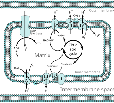
I mitokondrierne bliver vores mad omsat til energi i form af ATP. Dette sker via citronsyre cyklus og de fem mitokondrie komplekser. Basis viden om dette kan læses i wikipedia. Her ses citronsyre cyklus og de fem komplekser:


Figur fra Wikipedia: Oxidative phosphorylation
https://en.wikipedia.org/wiki/Oxidative_phosphorylation

I øverste venstre hjørne ses ATP synthase også kaldet complex V. Complex V sætter en fosfat-gruppe på ADP, så der dannes ATP. Complex V er yderligere beskrevet i Wikipedia: ATP synthase:  https://en.wikipedia.org/wiki/ATP_synthase

Professor Paul Fisher fra La Trobe Universitet har anvendt Seahorse teknologi til at undersøge mitokondrier fra ME patienter. Han har vist, at ved belastning arbejder complex V ineffektivt. Når cellerne ikke er belastede kan ATP produktionen følge med, men ved belastning går det galt. Effekten af complex V er nedsat med cirka 25% - mere hos de mest syge og mindre hos de mere raske ME patienter.

Complex V dysreguleringen medfører, at de andre mitokondrie komplekser arbejder mere for at prøve at kompensere. 

Professor Paul Fisher har fremlagt sine forskningsresultater i en præsentation:

Conclusions from Paul Fisher's "Specific Mitochondrial Respiratory Defects & Compensatory Changes in ME/CFS Patient Cells":
  • ATP synthesis by Complex V is less efficient in ME/CFS cells
  • Nonmitochondrial O2 consumption (metabolism) and mitochondrial respiratory capacity are elevated in ME/CFS cells
  • Respiration abnormalities correlate with disease state
  • Proteins involved in transporting molecules across the mitochondrial membrane and in mitochondrial fatty acid oxidation are upregulated in ME/CFS cells
  • TOR Complex I activity is elevated in ME/CFS lymphoblasts and could be responsible for upregulation of mitochondrial proteins
  • ME/CFS cells may be less able to respond to additional acute energy demands because they have already "turned up" their capacity as much as possible
  • Cause-effect relationships need to be determined


Her er link til præsentationen:
Specific Mitochondrial Respiratory Defects & Compensatory Changes in ME/CFS Patient Cells

Bloggen Health Rising har skrevet om præsentationen:
Emerging Insights #II: “The Cellular Equivalent of Chronic Fatigue” Found in ME/CFS

Artikel af Daniel Missailidis, Sarah Annesley og Paul Fisher
Pathological Mechanisms Underlying Myalgic Encephalomyelitis/Chronic Fatigue Syndrome
https://www.preprints.org/manuscript/201907.0196/v1

Hvis du vil vide mere om ATP synthase (complex V), så er her er review fra 2019:
ATP Synthase: Structure, Function and Inhibition

torsdag den 18. juli 2019

Betydning af acetyl-grupper for ME sygdomsmekanismen

ME patienter har dysreguleret glykolyse. McGregor et al (1) mener, at denne dysregulering medfører:

  1. nedsat dannelse af acetyl-grupper
  2. histone deacetylering
  3. nedsat acetylering af enzymer


McGregor et al artikel:
Post-Exertional Malaise Is Associated with Hypermetabolism, Hypoacetylation and Purine Metabolism Deregulation in ME/CFS Cases

McGregor holder foredrag om forskningen: An Omic Analysis of ME/CFS – an Assessment of Potential Mechanisms:
https://www.youtube.com/watch?v=UvGMbbyGtvg

Bloggen Health Rising har gennemgået forskningen:
Emerging Insights #1: McGregor’s Grand Conception of ME/CFS
https://www.healthrising.org/blog/2019/06/15/emerging-insights-mcgregor-metabolism-chronic-fatigue-glycolysis/


Nu følger lidt basisviden, om de begreber, der anvendes i artiklen/foredraget.

Acetyl-grupper

Acetat er saltet af eddikesyre. Den kemiske formel for acetat er  [CH3COO]  og den kemiske formel for en acetyl-gruppe er   CH3CO,  Når en acetyl-gruppe sættes på en organisk forbindelse kaldes den en acetylering (engelsk: acetylation). Deacetylering er når acetyl-gruppen fjernes. Mange forskellige enzymer kan påsætte/fjerne acetyl-grupper i forskellige biokemiske sammenhænge.

Histoner

Histoner er de spoler, hvorpå vores DNA er oprullet. Tilgængeligheden af DNAet styres ved at påsætte/fjerne acetylgrupper.

HAT: Histone acetyltransferase sætter acetyl-grupper på histonerne
HDAC: Histone deacetylase fjerner acetyl-grupper fra histonerne

Læs om dette i wikipedia: Histone acetylation and deacetylation


Acetyl-CoA

Acetyl-CoA er et energi-rigt molekyle, som kan anvendes som brændstof i TCA-cyklus, men det anvendes også til at donere acetyl-grupper til histonerne. Acetyl-CoA dannes ved nedbrydning af pyruvat.

Her er link til en god figur, der viser anvendelsen af acetyl-CoA. Det er figur 3 fra reference 2.
https://www.ncbi.nlm.nih.gov/pmc/articles/PMC5934764/figure/embr201745432-fig-0003/

Det tyder på, at pyruvat dehydrogenase komplekset kan rykke ind i cellens kerne og producere acetyl-grupper til histonerne: Se figur 4 fra reference 2.
https://www.ncbi.nlm.nih.gov/pmc/articles/PMC5934764/figure/embr201745432-fig-0004/


Mere viden

Og hvis man vil forske i emnet, så er der fine metoder til det:
Integrated Analysis of Acetyl-CoA and Histone Modification via Mass Spectrometry to Investigate Metabolically Driven Acetylation

Ovenstående artikel har et let forståelig figur:


Referencer

1) Mcgregor et al: Post-Exertional Malaise Is Associated with Hypermetabolism, Hypoacetylation and Purine Metabolism Deregulation in ME/CFS Cases Diagnostics (Basel). 2019 Jul 4;9(3). pii: E70. doi: 10.3390/diagnostics9030070. https://www.ncbi.nlm.nih.gov/pubmed/31277442

2) Lisowski et al: Mitochondria and the dynamic control of stem cell homeostasis.
EMBO Rep. 2018 May;19(5). pii: e45432. doi: 10.15252/embr.201745432. Epub 2018 Apr 16.
https://www.ncbi.nlm.nih.gov/pubmed/29661859


torsdag den 11. juli 2019

ME/SEID hypotese: Formindsket regeneration af dihydrolipinsyre

Der er kommet en ny ME/SEID hypotese (1):

Hypotesen går ud på, at der er formindsket regeneration af dihydrolipinsyre (dihydrolipoic acid) til lipinsyre (lipoic acid).

For at forstå hypotesen til bunds er det nødvendigt med basisviden om lipoic acid. Jeg vil derfor gennemgå basisviden fra wikipedia og fra artiklen:

Lipoic acid metabolism and mitochondrial redox regulation (2).

Jeg benytter de engelske ord for de kemiske forbindelser, så de er lettere at genfinde i wikipedia og i artiklen.

Lipoic acid (LA) og 2-keto-acid dehydrogenase komplekserne

Lipoic acid (LA) bliver dannet ud fra caprylic acid (= octanoic acid). Octa betyder otte. Dvs. LA dannes af en fedtsyre, der har en kulstof kæde-længde på otte kulstof atomer (C). Fedtsyrekæden har fået sat et svovl atom på kulstof atom nr. 6 og 8. De to svovl atomer er bundet sammen i en disulfid-binding (-S-S-).


Lipoic acid.svg

Figur er fra Wikipedia: Lipoic acid

Disulfid-bindingen i lipoic acid leverer reduktionspotentiale til de tre 2-keto-acid dehydrogenase komplekser:

  • PDC, pyruvate dehydrogenase complex
  • OGDC, 2-oxo-glutarate dehydrogenase comples (2-oxo-glutarate = alpha-ketoglutarate)
  • BCKDC, branched-chain alpha-keto dehydrogenase complex

Disse enzym komplekser er ens opbygget af tre subunits:

  • E1: decarboxylase
  • E2: lipoyltransferase
  • E3: dihydrolipoamide dehydrogense

PDC subunits:
E1: PDHA1,  pyruvate dehydrogenase E1 alpha 1 subunit
E2: DLATdihydrolipoamide acetyltransferase
E3. DLD,  dihydrolipoamide dehydrogenase
Herudover findes der et E3 bindings-protein (E3BP), som binder E2 og E3 sammen. E3BP kaldes også for PDC component X og kodes af genet PDHX

OGDC subunits:
E1: OGDH,  oxoglutarate dehydrogenase
E2: DLST, dihydrolipoamide S-succinyltransferase
E3. DLD,  dihydrolipoamide dehydrogenase

BCKDC subunits:
E1: BCKDHA,  branched chain keto acid dehydrogenase E1 subunit alpha og
BCKDHB branched chain keto acid dehydrogenase E1 subunit beta
E2: DBT,  dihydrolipoamide branched chain transacylase E2
E3. DLD,  dihydrolipoamide dehydrogenase

Som man kan se, benytter de tre enzym komplekser sammen E3 subunit: DLD. Herudover findes der et glycine cleavage system (GCS), som også benytter DLD.

GCS består af:
AMT, aminomethyltransferase
GLDC, glycine decarboxylase
DLD,  dihydrolipoamide dehydrogenase
GCSH, glycine cleavage system protein H

Der findes endvidere et enzym 2-oxoadipate dehydrogenase (2-OADH), som også kaldes dehydrogenase E1 and transketolase domain containing 1 ((DHTKD1). DHTKD1 decarboxylerer 2-oxoadipate til glutaryl-CoA, som sidste trin i nedbrydning af lysine. Enzymet indgår også i omsætning af  tryptophan.

Mitochondrial 2-oxoglutarate-dehydrogenase-complex-like protein består af:
DHTKD1, dehydrogenase E1 and transketolase domain containing 1
og så mener man, at enzymkomplekset deler E2 og E3 subunit med OGDC (3).

2-ketoacid dehydrogenaserne og deres betydning for TCA cyklus er vist i figur 3 i reference 2:
https://www.ncbi.nlm.nih.gov/pmc/articles/PMC5961061/figure/F3/

Nu standser vi op et øjeblik og tænker! Hypotesen siger: "Impairment of the E3 subunit...". Hvis hypotesen er sand på dette punkt, kan der forventes en påvirkning af alle enzym komplekser, der anvender E3 subunit DLD.  Det vil jeg vende tilbage til. Vi skal først forstå lipoic acid's rolle til bunds. 

Dannelse af lipoylated E2 subunit

Når lipoic acid bygges sammen med et protein, f.eks. enzymet subunit E2, kaldes det en lipoylation. Proteinet er blevet lipoylated. Den biokemiske stivej, der sørger for dette er ikke 100% klarlagt, så man møder en del uafklarede spørgsmål i litteraturen.

Figur 1B i reference 2 viser bedste bud på lipoylation af E2 subunit, som man mener, at det foregår hos mennesker.

Først dannes en octanoyl-ACP. ACP kaldes også for NDUFAB1 (ikke vist på figuren). Og så følger de tre trin beskrevet i figuren, hvor tre enzymer benyttes:

LIPT2lipoyl (octanoyl) transferase 2
LIAS,  lipoic acid synthetase
LIPT1,  lipoyltransferase 1

Structures, enzymes, and reaction mechanisms of lipoic acid metabolism (figur 1 fra reference 2).   https://www.ncbi.nlm.nih.gov/pmc/articles/PMC5961061/figure/F1/

Jeg vil i det følgende gennemgå figuren, men først forklarer jeg dannelsen af octanoyl-ACP.

Dannelse af octanoyl-ACP

Octanoic acid er en organisk syre. En organisk syre er kendetegnet ved at have en -COOH gruppe. Når -OH gruppen fjernes fra en organisk syre, så opstår en acyl, som er et mellemprodukt til en videre biokemisk reaktion. Man benytter bogstaverne -oyl i nanvnet på den opståede acyl-gruppe.Enkelte af de kemiske forbindelser kaldes dog for -yl, som f.eks. formyl og acetyl.

Når octanoic acid får fjernet sin OH gruppe bliver den til en octanoyl. Dette octanoyl mellemprodukt bæres rundt i cellen af et acyl carrier protein (ACP). Det hedder en octanoyl-ACP. Octanoyl og ACP er bundet sammen via et svovl atom (-S-). 

LIPT2 - første trin i dannelse af lipoylated E2 subunit: Octanoyl-H-protein dannes

LIPT2 er genet for enzymet lipoyl (octanoyl) transferase 2. Transfer bedtyder overfører. LIPT2 overfører octanoyl fra octanoyl-ACP til et protein H under fraspaltning af ACP. Hermed dannes octanoyl-H protein. (Protein H anvendes også i GCS komplekset). Octanoyl og H protein er bundet sammen med en amid binding (-NH-). Dette er illustreret i første trin i figur 1B i reference 2.

LIAS - andet trin i dannelse af lipoylated E2 subunit: Lipoylated H-protein dannes

LIAS er genet for enzymet lipoic acid synthetase. LIAS sætter to svovl atomer (S) på octanoyl-H proteinet. Hermed dannes lipoylated H protein. Dvs et protein koblet sammen med lipoic acid. LIAS anvender svovl-atom donation fra et (4Fe-4S) cluster og reduktiv spaltning af S-adenosylmethionine (SAM) for at danne 5'deoxyadenosyl-5'radikaler, der kan fjerne hydrogenatomer (H) fra kulstof atom (C) nr 6 og nr 8 og i stedet indsætte svovatomer (S). Dette er illustreret i andet trin i figur 1B fra reference 2.

LIPT1 - tredie trin i dannelse af lipoylated E2 subunit: Lipoylated E2 subunit  dannes

LIPT1 er genet for enzymet lipoyltransferase 1. LIPT1 fjerner H protein fra octanoyl og sætter i stedet E2 subunit på. Hermed dannes lipoylated E2 subunit. Dette er illustreret i tredie trin i figur 1B fra reference 2.

Af reference 4 figur 1 fremgår det, at lipoylated E2 subunit kan dannes UDEN først at anvende et H-protein:


Fig 1. Lipoic acid biosynthesis.
mtFAS generates octanoyl-ACP, that enters the lipoic acid biosynthesis pathway. The octanoyl moiety is then transferred from ACP to H or E2 proteins. Subsequently, insertion of two sulfur atoms occurs on the octanoyl moiety to generate lipoylated H or E2 proteins. 2-KGDH, α-ketoglutarate dehydrogenase; 2-OADH, 2-oxoadipate dehydrogenase; ACP, acyl carrier protein; BCKDH, branched-chain ketoacid dehydrogenase; GCS, glycine cleavage system; LA, lipoic acid; LIAS, lipoic acid synthetase; LIPT1, lipoyl(octanoyl) transferase 1; LIPT2, lipoyl(octanoyl) transferase 2; mtFAS, mitochondrial fatty acidynthesis; PDH, pyruvate dehydrogenase.

Dihydrolipoamide

E3 subunit hedder "dihydrolipoamide dehydrogenase. Dihdro betyder to hydrogen (H). Det betyder, at svovlbindingen i lipoic acid er brudt, og der er sat en H på hver S. Amid er en -NH2, der er sat på C=O, dvs en amid er det neutrale derivat af en carboxylsyre. Hvis det ene H fjernes, så har vi en amid-bindingsgruppe -NH-, som kan kobles på en E2 subunit. E3 subunit fjerner to H, dvs den dehydrogenerer dihydrolipoamide. Heraf navnet dihyrolipoamide dehydrogense.


Dihydrolipoamide.svg

Figur fra Wikipedia: Dihydrolipoamide


Den subunit-bundne lipoic acid og dihydrolipoic acid og deres skifte mellem (-S-S-) og (-SH) gør, at cellen kan flytte rundt på elektroner og protoner (H+). 

Flere figurer kan findes her "Redox reactions involving thiols and disulfides":
https://chem.libretexts.org/Bookshelves/Organic_Chemistry/Book%3A_Organic_Chemistry_with_a_Biological_Emphasis_(Soderberg)/16%3A_Oxidation_and_reduction_reactions/16.12%3A_Redox_reactions_involving_thiols_and_disulfides


Det skal fremhæves, at artiklen  påpeger, at kosttilskud af lipoic acid (LA) ikke hjælper: "The lack of an independent salvage pathway in humans abrogates the use of LA supplementation as a therapeutic option" (2).


2-keto-acid dehydrogenase komplekser i funktion

Figur 2 i reference 2 viser hvordan de tre subunits fungerer i samspil:
https://www.ncbi.nlm.nih.gov/pmc/articles/PMC5961061/figure/F2/

E1 benytter cofaktoren thiamine pyrophosphate  (TPP) til at decarboxylere alpha-keto acid. Herved dannes en acyl-TPP.
E2 overfører acyl-gruppen til Coenzym A (CoA). Herved dannes en acyl-CoA og den E2 bundne lipoic acid (LA) bliver til dihydrolipoamide (DHLA).
E3: Lipoic acid har under processen i E2 modtaget to hydrogen og er blevet til dihydrolipoamide (DHLA). Der er en sving-arm mellem E2 og E3, der flytter DHLA molekylet fra E2 til E3, I E3 fjernes de to hydrogen atomer og DHLA bliver herved regeneret til LA. Svingarmen kommer tilbage til E2 med LA. E3 benytter FAD og NAD til regenerering af DHLA til LA.

E3 subunit's kapacitet til at regenerere oxideret lipoic acid kontrolleres af NAD+/NADH ratio i mitokondrierne. Når der mangler NAD+, oxideres FADH2 i E3 subunit, og der dannes frie radikaler, som inaktiverer alpha-keto acid komplekserne.

PDC og OGDC kan beskyttes mod oxidativt stress ved glutathionylation. Gluathionylation inaktiverer enzymkomplekset. Glutathione-modifikationen kan igen fjernes med glutathione reductase 2, som kodes af genet GLRX2. OGDC bliver også beskyttet af thioredoxin reductase 2, som kodes af genet TXNRD2.

Figur 4 i reference to viser "Regulation of OGDH by reversible glutathionylation":
https://www.ncbi.nlm.nih.gov/pmc/articles/PMC5961061/figure/F4/


ME/SEID hypotese sammenholdt med ME forskningen

ME forskning der viser påvirkning af PDC

Nedsat PDC funktion hos ME patienter er allerede sandsynliggjort i Fluge, Mella et al's studie (5).

Det øgede lactat niveau er foreneligt med nedsat PDC funktion. Øget lactat niveau hos ME patienter i forbindelse med motion er for nylig dokumenteret af Lien et al (6).

ME forskning der viser påvirkning af PDHX

PDHX gene promoter var hypomethyleret i peripheral blood mononuclear blood cells (PBMC) fra ME patienter (7).

PDHX genekspression i PBMC ændres ved motion (8). McGregor et al (9) henviser til reference 8 i deres tabel S1, hvor opmærksomheden henledes på, at PDHX er reguleret af histone deacetylaserne HDAC1 og HDAC2. Histone regulering er påvirket hos ME patienter i forbindelse med post exertional malaise (PEM) (9).


ME forskning der viser påvirkning af DLAT (E2 subnit i PDC)

Sirtuin 4 (SIRT4) er et enzym med flere funktioner. SIRT4 kan fungere som E2 subunit lipomidase og klippe lipoamiden af E2 subuniten, og hermed nedregulere PDC aktivitet. Citat fra reference 10: "We determine that SIRT4 enzymatically hydrolyzes the lipoamide cofactors from the E2 component dihydrolipoyllysine acetyltransferase (DLAT), diminishing PDH activity. We demonstrate SIRT4-mediated regulation of DLAT lipoyl levels and PDH activity in cells and in vivo, in mouse liver."

Figur 1 fra reference 10: SIRT4 interacts with the pyruvate dehydrogenase complex
https://www.ncbi.nlm.nih.gov/pmc/articles/PMC4344121/figure/F1/

Der blev fundet øget mRNA ekspression af SIRT4 i PBMC fra ME patienter (p = 0,013) (5).

Der er blevet påvist spor af DLAT i spinalvæske fra ME patienter, men man ikke fandt DLAT spor hos kontrol personer eller hos Lyme patienter (patienter behandlet for borrelia infektion):
DLAT precursor, number of unique peptides identified in cerebrospinal fluid (table S1 in ref 11):
1) Controls: ingen værdi
2) ME patients: 1
3) Post treatment Lyme patients: ingen værdi


ME forskning der viser/ikke viser(?) påvirkning af DLD (den fælles E3 subnit)

Hvis DLD ikke fungerer kan man forvente, at alle DLD afhængige enzym komplekser er påvirkede. En genetisk mutation kan være dødelig (12): "E3-deficiency is a rare autosomal recessive genetic disorder frequently presenting with a neonatal onset and premature death."

Man må formode, at en mindre påvirkning af DLD vil give en mindre grad af alvorlighed. Spørgsmålet er således. Er alle DLD afhængige enzymer påvirkede hos ME patienter? Vi kender ikke svaret, men vi må holde øje med, hvad forskningen viser os.

En hæmning af OGDH vil give forhøjet niveau af alpha-ketoglutarate og 2-hydroxyglutarate, jvf figur 3 i reference 2. Et studie har vist forhøjet plasma niveau af alpha-ketoglutarate hos ME patienter (p = 0,003) (13). Gen ekspression af enzymet D-2-hydroxyglutarate dehydrogenase (D2HGDH) var opreguleret i fuldblod fra teen-age CFS patienter (p = 0,0555) (14). Genet D2HGDH var hypomethyleret (body) i PBMC fra ME patienter (15).

ME patienter anvender forgrenede aminosyrer som brændstof (5), så det må formodes at BCKDC fungerer.

Subunit E2 fra BCKDC hedder dihydrolipoamide branched chain transacylase E2 (DBT), og kaldes også for dihydrolipoyllysine-residue (2-methylpropanoyl) transferase. Plasma niveauet af S-(2-methylpropanoyl)-dihydrolipoamide var formindsket i plasma fra ME patienter (16). Hvad betyder dette fund?

Hvis glycine cleavage system (GCS) ikke fungerer, kommer der en ophobning af glycine. Glycine niveauet er normalt hos ME patienter. I reference 5 har ME pateinter et lidt højere serum niveau af glycine, men det er ikke statistisk signifikant (p = 0,082).

Gen promotor for AMT (som er den del af GCS) var hypomethyleret i PBMC fra ME patienter (7). Hvorfor?


ME forskning der viser påvirkning af mitokondrie fedtsyrer syntese - octanoic acid

Som det fremgår af reference 2, så starter produktionen af E2 subunit helt fra bunden af med en fedtsyre med 8 kulstofatmer, octanoic acid, (C8:0).

To uafhængig studier har påvist formindsket plasma niveau af 2-octenoylcarnitine (C8:1) hos ME patienter. Dvs carnitninen er sat sammen med en otte-kædet fedtsyre med en dobbeltbinding (17, 18).  Naviaux et al (17) kommenterer resultatet således: "Octenoic acid is produced as an intermediate of mitochondrial fatty acid and lipoic acid synthesis. It is the substrate for mitochondrial enoyl thioester reductase (ETR), a family of enzymes that requires NADPH to reduce the double bond to octanoic acid, which is then used for lipoic acid synthesis. Decreased levels of this C8:1 acylcarnitine are consistent with decreased mitochondrial fatty acid synthesis, increased oxidation, increased renal secretion, or a combination of the three."


ME forskning der viser påvirkning af mitokondrie fedtsyrer syntese - ACSM1

Der er diskussion om acyl-CoA synthetase medium chain family member 1, ACSM1, påvirker lipoic acid syntesen (2). I reference 2 står der: "There has been a report identifying a mammalian lipoic acid-activating enzyme that could activate exogenous lipoic acid (36); however, this function was ultimately attributed to the mitochondrial medium-chain acyl-CoA synthetase (ACSM1) (37, 38). This enzyme can utilize both the (R)- and (S)-enantiomers of LA and primarily uses GTP to activate the natural (R)-lipoic acid, but so far there has been no substantial evidence to support that this enzyme functions in LA metabolism in vivo (36). "

Om det er relevant eller ej skal det nævnes, at genet ACSM1 (TSS200) er påvist hypomethyleret i PBMC fra ME patienter i to uafhængige studier (7, 15). Ydermere er forskellig methylering af ACSM1 relateret til ME patient subtypes (19).


ME forskning der viser påvirkning af mitokondrie fedtsyrer syntese - ACACA og ACSF3

Malonyl-CoA er nødvendig for fedtsyre syntese i mitokondrierne og hermed også nødvendig for dannelse af lipoic acid. Acetyl-CoA carboxylase alpha (ACACA) katalyserer omdannelse af acetyl-CoA til malonyl-CoA. Acyl-CoA synthetase family member 3 (ACSF3) kan også danne malonyl-CoA. En mitokondriel isoform af ACACA og ACSF3 sørger i fællesskab for, at der er nok malonyl-CoA til at danne lipoic acid (20).

DNA methylering af ACACA og ACSF3 i PBMC fra ME patienter:

ACACA hypermethylering: 5'UTR;body og body (15), og body (21)

ACACA hypomethylering: to steder i 5'UTR, og to steder i en genic location, der ikke er angivet (7)

ACSF3 hypermethylering: tre steder body (15)

ACSF3 hypomethylering: tre steder 5'UTR og TSS100 (7), og body;5'UTR (15)

Forskellig methylering af ACSF3 (body) var relateret til ME patient subtypes (19).


ME forskning der viser påvirkning af LIPT1

Nogle mutationer i LIPT1 forårsager manglende aktivitet af PDC og OGDC, mens GCS stadig fungerer (22, 23). Der er rapporteret om dødelig lactis acidose i forbindelse med LIPT1 mutationer (24).

LIPT1 var hypermethyleret med en foldchange = 5,9 og p = 0,03 i CD4+ T celler fra ME patienter (25).


ME forskning der viser påvirkning af thioredoxin reductase og glutaredoxin 

TXNDR1 var hypomethyleret med en negativ foldchange = -3,6 og p = 0,000165 i CD4+ T celler fra ME patietner (25).

GLRX2 var hypermetyleret (TSS1500) i PBMC fra ME patienter i to studier (15, 21).

Gen ekspression af GLRX var opreguleret i fuldblod fra teen-age CFS patienter (p = 0,0005), hvilket var den laveste p-værdi i studiet (14).

Hvad sker der med redox i cellerne hos ME patienter? Og hvad betyder det for 2-keto-acid dehydrogenaser? Er de blevet glutathionylated?

Figur med Glutaredoxin (a) and thioredoxin (b) systems:
https://www.nature.com/articles/4401818/figures/1

Mere viden: Thioredoxin and glutaredoxin-mediated redox regulation of ribonucleotide reductase

Videre inspiration

Tak til Victoria Bohne, Oeyvind Bohne og de personer, der donerede til illustration af deres artikel om ME/SEID hypotesen. 

Vi skal helt sikkert et spadestik dybere ned i hvad der foregår i pyruvat dehydrogenase komplekset. Videre læsning må være:

Structural and Functional Analyses of the Human PDH Complex Suggest a “Division-of-Labor” Mechanism by Local E1 and E3 Clusters


Highlights herfra:

Binding of CoA to E2 primes PDHc lipoyl arms for subsequent E2 and E3 reactions


Referencer: 

1) Victoria J. Berdikova Bohne og Oeyvind BohneSuggested Pathology of Systemic Exertion Intolerance Disease: Impairment of the E3 Subunit or Crossover of Swinging Arms of the E2 Subunit of the Pyruvate Dehydrogenase Complex Decreases Regeneration of Cofactor Dihydrolipoic Acid of the E2 Subunit 
https://www.sciencedirect.com/science/article/abs/pii/S0306987718312994

2) Solomonson og DeBerardinis: Lipoic acid metabolism and mitochondrila redox regulation. J Biol Chem. 2018 May 18;293(20):7522-7530. doi: 10.1074/jbc.TM117.000259. Epub 2017 Nov 30.
https://www.ncbi.nlm.nih.gov/pubmed/29191830

3) Nimeria et al: The mitochondrial 2-oxoadipate and 2-oxoglutarate dehydrogenase complexes share their E2 and E3 components for their function and both generate reactive oxygen species Free Radic Biol Med. 2018 Feb 1;115:136-145. doi: 10.1016/j.freeradbiomed.2017.11.018. Epub 2017 Dec 1.
https://www.ncbi.nlm.nih.gov/pubmed/29191460

4) Bernadinelle et al: Mis-targeting of the mitochondrial protein LIPT2 leads to apoptotic cell death Plos One, june 9, 2017. https://journals.plos.org/plosone/article?id=10.1371/journal.pone.0179591

5) Fluge et al: Metabolic profiling indicates impaired pyruvate dehydrogenase function in myalgic encephalopathy / chronic fatigue syndrome. JCI Insight. 2016; 1(21):e89376. Doi 10.1172/jci.insight.89276

6) Lien et al: Abnormal blood lactate accumulation during repeated exercise testing in myalgic encephalomyelitis/chronic fatigue syndrome Physiol Rep. 2019 Jun;7(11):e14138. doi: 10.14814/phy2.14138.  https://www.ncbi.nlm.nih.gov/pubmed/31161646

7) Trivedi et al: Identification of ME/CFS - associated DNA methylation patterns. Plos One 2018, 13, 7 https://journals.plos.org/plosone/article?id=10.1371/journal.pone.0201066

8) Whistler et al: Exercise responsive genes measured in peripheral blood of women with chronic fatigue syndrome and matched control subjects BMC Physiol. 2005 Mar 24;5(1):5.

9) Mcgregor et al: Post-Exertional Malaise Is Associated with Hypermetabolism, Hypoacetylation and Purine Metabolism Deregulation in ME/CFS Cases Diagnostics (Basel). 2019 Jul 4;9(3). pii: E70. doi: 10.3390/diagnostics9030070. https://www.ncbi.nlm.nih.gov/pubmed/31277442

10) Mathias et al: Sirtuin 4 is a lipoamidase regulating pyruvate dehydrogenase complex activity. Cell. 2014 Dec 18;159(7):1615-25. doi: 10.1016/j.cell.2014.11.046. https://www.ncbi.nlm.nih.gov/pubmed/25525879

11) Schutzer et al: Distinct Cerebrospinal Fluid Proteomes Differentiate Post- Treatment Lyme Disease from Chronic Fatigue Syndrome. PLOS One February 2011, volume 6, Issue https://journals.plos.org/plosone/article?id=10.1371/journal.pone.0017287

12) Ambrus og Adam-Vizi: Human dihydrolipoamide dehydrogenase (E3) deficiency: Novel insights into the structural basis and molecular pathomechanism Neurochem Int. 2018 Jul;117:5-14. doi: 10.1016/j.neuint.2017.05.018. Epub 2017 Jun 2. https://www.ncbi.nlm.nih.gov/pubmed/28579060

13) Germain et al: Prospective Biomarkers from Plasma Metabolomics of Myalgic Encephalomyelitis/Chronic Fatigue Syndrome Implicate Redox Imbalance in Disease Symptomatology. Metabolites. 2018 Dec 6;8(4). pii: E90. doi: 10.3390/metabo8040090.
https://www.ncbi.nlm.nih.gov/pubmed/30563204

14) Nguyen et al: Whole blood gene expression in adolescent CFS: an exploratory crosssectional study suggesting altered B cell differentiation and survival. J Transl Med. 2017,15,102. https://www.ncbi.nlm.nih.gov/pubmed/28494812

15) de Vega et al: Epigenetic modifications and glucocorticoid sensitivity in ME/CFS. BMC Medical Genomics, 2017, 10, 11 https://www.ncbi.nlm.nih.gov/pmc/articles/PMC5324230/

16) Germain et al: Metabolic profiling of a ME/CFS discovery cohort reveals disturbances in fatty acid and lipid metabolism. Mol. BioSyst. 2017, 13, 371 https://pubs.rsc.org/en/Content/ArticleLanding/2017/MB/C6MB00600K#!divAbstract

17) Naviaux RK, Naviaux JC, Li K, Bright AT, Alaynick WA, Wang L, Baxter A, Nathan N et al (2016) Metabolic features of chronic fatigue syndrome. Proc Natl Acad Sci U S A 113:E5472–E5480. https://doi.org/10.1073/pnas.1607571113

18) Reuter and Evans: Long-chain acylcarnitine deficiency in patients with CFS. Potential involvement of altered carnitine palmitoyltransferase-I-activity. J. Int. Med. 2011, 270.

19) de Vega et al: Integration of DNA methylation & health scores identifies subtypes in ME/CFS. Epigenomics 2018, 10, 5 https://www.futuremedicine.com/doi/full/10.2217/epi-2017-015

20) Monteuuis et al. A conserved mammalian mitochondrial isoform of acetyl-CoA carboxylase ACC1 provides the malonyl-CoA essential for mitochondrial biogenesis in tandem with ACSF3. Biochem J. 2017 Nov 6;474(22):3783-3797. doi: 10.1042/BCJ20170416. https://www.ncbi.nlm.nih.gov/pubmed/28986507

21) de Vega et al: DNA methylation Modifications associated with CFS. PlosOne, 2014, 9, 8. https://journals.plos.org/plosone/article?id=10.1371/journal.pone.0104757

22) Soreze et al. Mutations in human lipoyltransferase gene LIPT1 cause a Leigh disease with secondary deficiency for pyruvate and alpha-ketoglutarate dehydrogenase Orphanet J Rare Dis. 2013 Dec 17;8:192. doi: 10.1186/1750-1172-8-192. https://www.ncbi.nlm.nih.gov/pubmed/24341803

23) Tort et al. Mutations in the lipoyltransferase LIPT1 gene cause a fatal disease associated with a specific lipoylation defect of the 2-ketoacid dehydrogenase complexes. Hum Mol Genet. 2014 Apr 1;23(7):1907-15. doi: 10.1093/hmg/ddt585. Epub 2013 Nov 20.https://www.ncbi.nlm.nih.gov/pubmed/24256811

24) Tache et al: Lipoyltransferase 1 Gene Defect Resulting in Fatal Lactic Acidosis in Two Siblings. Case Rep Obstet Gynecol. 2016;2016:6520148. doi: 10.1155/2016/6520148. Epub 2016 May 10. https://www.ncbi.nlm.nih.gov/pubmed/27247813


25) Brenu et al: Methylation profile of CD4+ T cells in CFS/ME. J. Clin Cell Immunol 5, 228https://www.omicsonline.org/open-access/methylation-profile-of-cd-t-cells-in-chronic-fatigue-syndromemyalgic-encephalomyelitis-2155-9899.1000228.php?aid=27598

mandag den 1. juli 2019

Mutations in the IDO2 gene and DNA methylations in genes in the NAD/NADP synthesis pathway in ME

Results shown at the NIH ME/CFS conference indicate that all of the 20 of the severely ill ME patients had damaging mutations in their indolamine 2,3-dioxygenase 2 (IDO2) gene (1,2).

The enzymes IDO1 and IDO2 catalyze the conversion of tryptophan to N-formyl-kynurenine. This is the first step in the kynurenine pathway and in the de novo pathway of biosynthesis of  NAD+  and  NADP+  (3).

Use link below to see "Biosynthesis of NAD(P)+ in mammalian cells" (3):
https://www.ncbi.nlm.nih.gov/pmc/articles/PMC5737637/figure/f2/

NAD+ is synthesized by three pathways: 

  • the de novo pathway
  • the Preiss–Handler pathway
  • the salvage pathway 


All of the enzymes in these three pathways are thoroughly described in reference 3. Some of these enzymes show up in ME research:

NAMPT = nicotinamide phosphoribosyltransferase. NAMPT is an enzyme in the salvage pathway.
NADSYN1 = NAD synthetase 1. NADSYN1 is an enzyme in the Preiss–Handler pathway.
QPRT = quinolinate phosphoribosyltransferase. QPRT is an enzyme the de novo pathway.
NADK = NAD kinase. NADK catalyzes the transfer of a phosphate group from ATP to NAD to generate NADP.

NAMPT gene expression was increased in whole blood from adolescent ME/CFS patients (adjusted p-value = 0,0621) (4).

NAMPT gene expression was increased in peripheral blood mononuclear cells (PBMC) from ME patients (5).


DNA methylations of genes in PBMC from ME patients:
  • NADSYN1 hypermethylated (gene probe cg 03146219) (6)
  • NADSYN1 hypomethylated (5'UTR) (7)
  • QPRT differentially methylated (1stExon) in ME patient subtypes (8)
  • NADK hypermethylated (gen probe cg00343906) (6)
  • NADK hypermethyltated (body) (9)
  • NADK differentially methylated (body) in ME patient subtypes (8)
  • NADK hypometylated (TSS1500) (7)


Kynurenine Is a Cerebrospinal Fluid Biomarker for Bacterial and Viral Central Nervous System Infections

The tryptophan-kynurenine-nicotinamide adenine dinucleotide (oxidized; NAD+) pathway is closely associated with regulation of immune cells toward less inflammatory phenotypes and may exert neuroprotective effects. Investigating its regulation in central nervous system (CNS) infections would improve our understanding of pathophysiology and end-organ damage, and, furthermore, open doors to its evaluation as a source of diagnostic and/or prognostic biomarkers (10).

The Trp-Kyn-NAD+ pathway is activated in CNS infections and provides highly accurate CSF biomarkers, particularly when combined with standard CSF indices of neuroinflammation (10).

The ME metabolic trap hypothesis predicts dysregulated Trp-Kyn- pathway in immune cells from ME patients (11).


Is the Trp-Kyn-NAD+ pathway dysregulated in the cerebrospinal fluid from ME patients?


References

1) National Institute of Health ME/CFS konference april 2019,
del 1: https://videocast.nih.gov/summary.asp?live=31636&bhcp=1
del 2: https://videocast.nih.gov/summary.asp?Live=31640&bhcp=1

2)  Health Rising,   NIH ME/CFS Conferencen, april 2019:
https://www.healthrising.org/blog/2019/04/10/nih-accelerating-chronic-fatigue-exhaustion-inflammation/

3) Xiao et al: NAD(H) and NADP(H) Redox Couples and Cellular Energy Metabolism Antioxid Redox Signal. 2018 Jan 20;28(3):251-272. doi: 10.1089/ars.2017.7216. Epub 2017 Jul 28.
https://www.ncbi.nlm.nih.gov/pubmed/28648096

4) Nguyen et al: Whole blood gene expression in adolescent CFS: an exploratory crosssectional study suggesting altered B cell differentiation and survival. J Transl Med. 2017,15,102.  https://www.ncbi.nlm.nih.gov/pubmed/28494812

5) 12) Sweetman et al: Changes in the transcriptome of circulating immune cells of a New Zealand cohort with myalgic encephalomyelitis/chronic fatigue syndrome. Sweetman et al: Int J Immunopathol Pharmacol. 2019 Jan-Dec;33:2058738418820402. doi: 10.1177/2058738418820402.
https://www.ncbi.nlm.nih.gov/pubmed/30791746

6) 4) de Vega et al: DNA methylation Modifications associated with CFS. PlosOne, 2014, 9, 8. https://journals.plos.org/plosone/article?id=10.1371/journal.pone.0104757

7) Trivedi et al: Identification of ME/CFS - associated DNA methylation patterns. Plos One 2018, 13, 7 https://journals.plos.org/plosone/article?id=10.1371/journal.pone.0201066

8) de Vega et al: Integration of DNA methylation & health scores identifies subtypes in ME/CFS. Epigenomics 2018, 10, 5 https://www.futuremedicine.com/doi/full/10.2217/epi-2017-015

9) de Vega et al: Epigenetic modifications and glucocorticoid sensitivity in ME/CFS. BMC Medical Genomics, 2017, 10, 11 https://www.ncbi.nlm.nih.gov/pmc/articles/PMC5324230

10) Sühs et al: Kynurenine Is a Cerebrospinal Fluid Biomarker for Bacterial and Viral Central Nervous System Infections J Infect Dis. 2019 Jun 5;220(1):127-138. doi: 10.1093/infdis/jiz048.
https://www.ncbi.nlm.nih.gov/pubmed/30721966

11) Metabolic Traps in ME/CFS - Research Update by Dr. Robert Phairhttps://www.youtube.com/watch?v=Quh-77gvw4Q