Sider

mandag den 9. september 2019

Genomics and the cytokine network in ME

Genomics in ME

Gene variants of IL12B, IL1B and IL4R have been identified as a risk loci in ME (1):

image.png

Slide from: Whole Genome Sequencing and Analysis of ME/CFS
https://www.youtube.com/watch?v=nIJX-Q7w_Z4


Cytokines in ME

Plasma cytokine levels have been measured in ME patients with short-duration illness (≤3 years) and ME patients with long-duration illness (>3 years) (2).

IL-12B (IL-12p40) was increased in short-duration and decreased in long-duration compared to normal controls.

IL-1β was decreased in long-duration compared to short-duration and normal controls (2).

IL4 was increased in short-duration and decreased in long-duration compared to normal controls (2).

Interferon-γ (IFNγ) was decreased in long-duration compared to short-duration and normal controls (2).


Cytokines and IDO1

IL-12B stimulate the production of IFNγ, and  IFNγ induce IDO1 activity (3).

IDO1 is inhibited by IL-4 (3).

Do the risk loci play a role in the ME-IDO-metabolic trap hypothesis?


Further reading:

Genomics, proteomics and transcriptomics show the iNOS pathway is upregulated in ME
http://followmeindenmark.blogspot.com/2019/09/genomics-proteomics-and-transcriptomics.html


References:

1) Whole Genome Sequencing and Analysis of ME/CFS https://www.youtube.com/watch?v=nIJX-Q7w_Z4

2) Hornig et al: Distinct plasma immune signatures in ME/CFS are present early in the course of illness. Sci Adv. 2015 Feb;1(1). pii: e1400121.  https://www.ncbi.nlm.nih.gov/pubmed/26079000

3) Opitz et al: Tryptophan degradation in autoimmune diseases. Cell Mol Life Sci. 2007 Oct;64(19-20):2542-63. https://www.ncbi.nlm.nih.gov/pubmed/17611712

søndag den 1. september 2019

Genomics, proteomics and transcriptomics show the iNOS pathway is upregulated in ME

Activated macrophages produce an inducible NO synthase (iNOS or NOS2).  Although iNOS was originally identified and characterized in macrophages, it is present in numerous cell types including endothelial cells, fibroblasts, vascular smooth muscle cells and cardiac myocytes (1).


Genomics in ME

A gene variant of iNOS has been identified as a risk locus in ME (2):

image.png

Slide from. Whole Genome Sequencing and Analysis of ME/CFS
https://www.youtube.com/watch?v=nIJX-Q7w_Z4 


Proteomics in ME

The pathway "Production of NO and ROS in macrophages" was upregulated in the cerebrospinal fluid from ME patients (table S6 in ref 3).


Transcriptomics in ME

Functional Network Analysys of gene transcripts from ME immune cells showed thar the pathway "Production of NO and ROS in macrophages" was enriched (p-value = 0,000018) (4).


Macrophages and the ME-IDO-metabolic trap

The ME-IDO metabolic trap hypothesis tell us that the kynurenic pathway may be blocked in dendritic cells and macrophages. This means decreased metabolites from the pathway (5).

During inflammation and resolution of inflammation, this may be going on in normal cells (6) :
  • Increased IDO activity
  • Increased kynurenic acid (KYNA) production
  • Increased GPR35 activation (KYNA activates GPR35)
  • Decreased iNOS expression


Figure 2 from ref 6 Wirthgen et al (2018).


Is this what is going on in ME macrophages???:
  • Decreased IDO activity
  • Decreased KYNA production
  • Decreased GPR35 activation
  • Increased iNOS expression


References:

1) iNOS signaling: https://www.qiagen.com/us/shop/genes-and-pathways/pathway-details/?pwid=252

2) Whole Genome Sequencing and Analysis of ME/CFS https://www.youtube.com/watch?v=nIJX-Q7w_Z4

3) Schutzer et al: Distinct Cerebrospinal Fluid Proteomes Differentiate Post- Treatment Lyme Disease from Chronic Fatigue Syndrome. PLOS One February 2011, volume 6, Issuehttps://journals.plos.org/plosone/article?id=10.1371/journal.pone.0017287

4) Sweetman et al: Changes in the transcriptome of circulating immune cells of a New Zealand cohort with myalgic encephalomyelitis/chronic fatigue syndrome. Sweetman et al: Int J Immunopathol Pharmacol. 2019 Jan-Dec;33:2058738418820402. doi: 10.1177/2058738418820402.
https://www.ncbi.nlm.nih.gov/pubmed/30791746

5) Kashi AA Davis RW and, Phair RD: The IDO Metabolic Trap Hypothesis for the Etiology of ME/CFS. Diagnostics (Basel). 2019 Jul 26;9(3). pii: E82. doi: 10.3390/diagnostics9030082. https://www.mdpi.com/2075-4418/9/3/82

Metabolic Traps in ME/CFS - Research Update by Dr. Robert Phair
https://www.youtube.com/watch?v=Quh-77gvw4Q

6) Wirthgen E Hoeflich A, Rebl A, Günther J.: Kynurenic Acid: The Janus-Faced Role of an Immunomodulatory Tryptophan Metabolite and Its Link to Pathological Conditions
Front Immunol. 2018 Jan 10;8:1957. doi: 10.3389/fimmu.2017.01957. eCollection 2017.
https://www.frontiersin.org/articles/10.3389/fimmu.2017.01957/full

søndag den 25. august 2019

Does OXA1L, MRM2 and MRRF protein activity compensate for complex V inefficiency in ME patient cells?

ATP synthesis by Complex V is less efficient in ME/CFS cells. The other mitochondrial complexes work harder to compensate (1).

Is this problem reflected in previous ME research?


OXA1L

Oxidase (cytochrome c) assembly 1-like (OXA1L) is a mitochondrial inner membrane protein. It is required for the insertion of integral membrane proteins into the mitochondrial inner membrane. Essential for the activity and assembly of cytochrome oxidase. Required for the correct biogenesis of complex V and complex I in mitochondria (2).

Knockdown of human Oxa1L impairs the biogenesis of complex V and NADH:ubiquinone oxidoreductase (3).

The C-terminal approximately 100-amino acid tail of Oxa1L (Oxa1L-CTT) binds to mitochondrial ribosomes and plays a role in the co-translational insertion of mitochondria-synthesized proteins into the inner membrane (4).

The gene OXA1L had changed DNA methylation profile and increased foldchange (2,01) in CD4+ T-cells from ME patients (5).


MRM2/FTSJ2

Mitochondrial rRNA methyltransferase 2 (also known as FTSJ2) is involved in mitoribosome assembly (6).

Defective MRM2 causes MELAS-like clinical syndrome (MELAS = mitochondrial encephalopathy, lactic acidosis and stroke-like episodes (7).

The gene MRM2 had changed DNA methylation profile and increased foldchange (2,12) in CD4+ T cells from ME patients (5).

The gene MRM2 was hypomethylated (3'UTR) in peripheral mononuclear cells (PBMC) from ME patients (8).


MRRF

Mitochondrial ribosome-recycling factor (MRRF) is responsible for the release of ribosomes from messenger RNA at the termination of protein biosynthesis. May increase the efficiency of translation by recycling ribosomes from one round of translation to another (2).

MRRF is essentiel for cell viability and depletion of MRRF leads to loss of mitochondrial complexes (9).

MRRF gene expresseion was increased in PBMC from ME patients (10, 11). 


Does OXA1L, MRM2 and MRRF protein activity compensate for complex V inefficiency in ME patient cells?


References

1) Missailidis, D.; Annesley, S.J.; Allan, C.Y.; Sanislav, O.; Lidbury, B.A.; Lewis, D.P.; Fisher, P.R. An isolated Complex V defect and dysregulated mitochondrial function in immortalized lymphocytes from ME/CFS patients. Submitted 2019.

Specific Mitochondrial Respiratory Defects & Compensatory Changes in ME/CFS Patient Cells
https://www.youtube.com/watch?v=SjK39QCPeeY


3) Stiburek et al. Knockdown of human Oxa1l impairs the biogenesis of F1Fo-ATP synthase and NADH:ubiquinone oxidoreductase. J Mol Biol. 2007 Nov 23;374(2):506-16. Epub 2007 Sep 20. https://www.ncbi.nlm.nih.gov/pubmed/17936786

4) Hauge et al: Properties of the C-terminal tail of human mitochondrial inner membrane protein Oxa1L and its interactions with mammalian mitochondrial ribosomes. J Biol Chem. 2010 Sep 3;285(36):28353-62. doi: 10.1074/jbc.M110.148262. Epub 2010 Jul 2. https://www.ncbi.nlm.nih.gov/pubmed/20601428

5) Brenu et al: Methylation profile of CD4+ T cells in CFS/ME. J. Clin Cell Immunol 5, 228https://www.omicsonline.org/open-access/methylation-profile-of-cd-t-cells-in-chronic-fatigue-syndromemyalgic-encephalomyelitis-2155-9899.1000228.php?aid=27598

6) Rorbach et al: MRM2 and MRM3 are involved in biogenesis of the large subunit of the mitochondrial ribosome. Mol Biol Cell, 2014, 25, 17.

7) Garone et al: Defective MRM2 causes MELAS-like clinical syndrome. Hum Mol Genet, 2017, 26, 21.

8) Trivedi et al: Identification of ME/CFS - associated DNA methylation patterns. Plos One 2018, 13, 7.

9) Rorbach et al: The human mitochondrial ribosome recycling factor is essential for cell viability. Nucleic Acids Research, 2008, 36, 18.

10) Kerr et al: Gene expression subtypes in patients with CFS/ME. JID, 2008, 197.

11) Frampton et al: Assessment of a 44 gene classifier for the evaluation of CFS from PBMC gene expression. Plos one, 2011, 6, 3.

torsdag den 22. august 2019

AMPD3 in ME

Adenosine monophosphate deaminase (AMPD) converts AMP to inosine monophosphate (IMP).

The AMP deaminase gene family:
AMPD1, muscle (m) isoform
AMPD2, liver (l) cells isoform
AMPD3, erothrocyte (e) isoform

The isoforms are named after their predominant location, but are also expressed in other tissue. Fx. AMPD3 is also expressed in muscles.

The purine nucleotide cycle of muscles consist of the conversion of AMP→IMP→AMP and requires AMP deaminase. Flux through this cycle increases during exercise.


Genomics in ME

A gene variant of AMPD3 has been identified as a risk locus in ME (1):

image.png

Slide from. Whole Genome Sequencing and Analysis of ME/CFS
The genes AMPD2 and AMPD3 were epigenetic changed in peripheral blood mononuclear cells (PBMC) from ME patients in two studies (2, 3). Some of the DNA methylations in AMPD2 and AMPD3 were related to quality of life in the ME patients (3). AMPD2 and AMPD3 were differntially methylated in PBMC from ME patient subtypes (4).


Transcriptomics in ME

AMPD3 gene expression was downregulated in biopsies from the vastus lateralis muscle in ME patients (5).


Metabolomics in ME

Metabolic profiling of ME patients showed disturbances in purine metabolism (6, 7, 8). Fx, IMP was decreased in plasma from ME patients (8). 


HDAC3 and AMPD3

Depletion of histone deacetylase 3 (HDAC3) in skeletal muscle in mice causes lower glucose utilization and greater lipid oxidation. AMPD3 is involved in the regulation of the fuel switch (9).

Interestingly, very low density lipoprotein receptor (VLDLR) was found to be up-regulated in the ME muscle biopsies in ref 5. VLDLR is responsible for VLDL uptake into the fiber and is involved in the primary pathway of fatty acid transport in skeletal muscle (5). 

6-phosphofructo-2-kinase/fructose-2,6-biphosphatase 3 (PFKFB3) was down-regulated (5). PFKFB3 belongs to a family of bifunctional proteins that are involved in both the synthesis and degradation of fructose-2,6-bisphosphate, a regulatory molecule that controls glycolysis.

Transducin beta like 1 X-linked receptor 1 (TBL1XR1) was up-regulatede in women and down-regulted in men (5). TBL1XR1 is thought to be a component of both nuclear receptor corepressor (N-CoR) and HDAC3 complexes.

ME is associated with purine and histone deacetylation dysregulation (10). 


References:

1) Whole Genome Sequencing and Analysis of ME/CFS https://www.youtube.com/watch?v=nIJX-Q7w_Z4

2) ) de Vega et al: Epigenetic modifications and glucocorticoid sensitivity in ME/CFS. BMC Medical Genomics, 2017, 10, 11 https://www.ncbi.nlm.nih.gov/pmc/articles/PMC5324230/

3) Trivedi et al: Identification of ME/CFS - associated DNA methylation patterns. Plos One 2018, 13, 7 https://journals.plos.org/plosone/article?id=10.1371/journal.pone.0201066

4) de Vega et al: Integration of DNA methylation & health scores identifies subtypes in ME/CFS. Epigenomics 2018, 10, 5 https://www.futuremedicine.com/doi/full/10.2217/epi-2017-015

5) Pietrangelo et al: Transcription profile analysis of vastus lateralis muscle from patients with chronic fatigue syndrome. Int J Immunopathol Pharmacol. 2009 Jul-Sep;22(3):795-807. https://www.ncbi.nlm.nih.gov/pubmed/19822097

6) Naviaux RK, Naviaux JC, Li K, Bright AT, Alaynick WA, Wang L, Baxter A, Nathan N et al (2016) Metabolic features of chronic fatigue syndrome. Proc Natl Acad Sci U S A 113:E5472–E5480. https://doi.org/10.1073/pnas.1607571113

7) Germain et al: Metabolic profiling of a ME/CFS discovery cohort reveals disturbances in fatty acid and lipid metabolism. Mol. BioSyst. 2017, 13, 371 https://pubs.rsc.org/en/Content/ArticleLanding/2017/MB/C6MB00600K#!divAbstract

8) Germain et al: Prospective Biomarkers from Plasma Metabolomics of Myalgic Encephalomyelitis/Chronic Fatigue Syndrome Implicate Redox Imbalance in Disease Symptomatology. Metabolites. 2018 Dec 6;8(4). pii: E90. doi: 10.3390/metabo8040090.
https://www.ncbi.nlm.nih.gov/pubmed/30563204

9) Hong et al: Dissociation of muscle insulin sensitivity from exercise endurance in mice by HDAC3 depletion. Nat Med. 2017 Feb;23(2):223-234. doi: 10.1038/nm.4245. Epub 2016 Dec 19. https://www.ncbi.nlm.nih.gov/pubmed/27991918

10) Mcgregor et al: Post-Exertional Malaise Is Associated with Hypermetabolism, Hypoacetylation and Purine Metabolism Deregulation in ME/CFS Cases Diagnostics (Basel). 2019 Jul 4;9(3). pii: E70. doi: 10.3390/diagnostics9030070. https://www.ncbi.nlm.nih.gov/pubmed/31277442

fredag den 26. juli 2019

Complex V is down in ME - does it also explain Electromagnetic Hypersensitivity?

Complex V

Complex V (also known as ATP synthase or F0F1ATPase) is working less efficiently in cells from ME patients (1).


Chemical Intolerance and Electromagnetic Hypersensitivity in ME

ME patients have chemical intolerance (CI), also knowns as multiple chemical sensitivity (MCS). 

I have asked the question:
Complex V is down in ME - does it explain Chemical Intolerance?
http://followmeindenmark.blogspot.com/2019/07/complex-v-is-down-in-me-does-it-explain.html

Several ME patients also have electromagnetic hypersensitivity (EHS). It has been hypothesized that EHS and CI are two etiopathogenic aspects of a unique pathological disorder (2, 3).

Maybe ME, CI and EHS are a unique "Complex V-disease"?


Electromagnetic Fields and Complex V

Humans are exposed to electromagnetic fields (EMFs) from: Radio and television stations and receivers, radar, computers, Wi-Fi antennas, mobile phones, microwave ovens, and many devices used in medicine and industry. Our blood is just under the skin and is exposed to EMFs all the time.

Lassaivia et al have investigated peripheral blood lympho-monocytes exposed to EMFs at 1.8 GHz frequency and 200 V/m electric field strength. Respirometric measurements of mitochondrial activity in intact lympho-monocytes showed:
  • An in increase of the resting oxygen consumption rate after 20 h of exposure, which was coupled to a significant increase of the FoF1-ATP synthase-related oxygen consumption.
  • At lower time-intervals of EMFs exposure (i.e. 5 and 12 h) a large increase of the proton leak-related respiration was observed which, however, recovered at control levels after 20 h exposure.
  • No significant variations in the mitochondrial mass/morphology was observed in EMFs-exposed lympho-monocytes.
  • Altered redox homeostasis was shown in EMFs-exposed lympho-monocytes, which progressed differently in nucleated cellular subsets.

The results suggest the occurrence of adaptive mechanisms put in action, likely via redox signaling, to compensate for early impairments of the oxidative phosphorylation system caused by exposure to EMFs (4).


ME and Complex V

Fisher et al have investigated lymphoblasts from ME patients and found specific mitochondrial respiratory defects and compensatory changes (5).

Respirometry of immortalized ME patient lymphocytes (lymphoblasts) showed:
  • Mitochondrial ATP synthesis by Complex V is inefficient, representing a significantly lower proportion of the basal mitochondrial respiratory activity.
  • Absolute ATP synthesis rates (pmol/min) were not significantly lower than in control cells, while glycolysis rates and steady state ATP levels were unchanged.
  • There was a significant increases in maximum respiratory capacity, including Complex I activity.
  • “Nonmitochondrial” O2 consumption by other cellular enzymes was also elevated. 

The results suggest that ME/CFS cells compensate for the reduced efficiency of ATP synthesis by upregulating mitochondrial respiratory capacity.

The mitochondrial membrane “mass” and genome copy number were unchanged. Thus ME/CFS cells do not have “more” mitochondria, but their mitochondria have greater respiratory capacity. This increased capacity is underutilized because of the Complex V defect, so that the respiratory “spare capacity” was increased and the mitochondrial membrane potential was elevated (5).

I have a new question:
Can continous exposure from electromagnetic fields in the environment put further strain on a compromised complex V in ME patient cells?


A magnetic field is able to influence Complex V

Interestingly, a team of Japanese scientists have succeeded in attaching magnetic beads to the stalks of F1 -ATPase isolated in vitro, which rotated in presence of a rotating magnetic field. F1 -ATPase synthesized ATP from ADP and Pi when rotated in a clockwise direction at a rate of about 5 molecules per second. Additionally, ATP was hydrolyzed when the stalks were rotated in the counterclockwise direction or when they were not rotated at all (6, ref 26 in ref 7).


References

1) Missailidis, D.; Annesley, S.J.; Allan, C.Y.; Sanislav, O.; Lidbury, B.A.; Lewis, D.P.; Fisher, P.R. An isolated Complex V defect and dysregulated mitochondrial function in immortalized lymphocytes from ME/CFS patients. Submitted 2019.

Specific Mitochondrial Respiratory Defects & Compensatory Changes in ME/CFS Patient Cells
https://www.youtube.com/watch?v=SjK39QCPeeY

2) Belpomme D, Campagnac C, Irigaray P. : Reliable disease biomarkers characterizing and identifying electrohypersensitivity and multiple chemical sensitivity as two etiopathogenic aspects of a unique pathological disorder. Rev Environ Health. 2015;30(4):251-71. doi: 10.1515/reveh-2015-0027. https://www.ncbi.nlm.nih.gov/pubmed/26613326

3) De Luca C, Thai JC, Raskovic D, Cesareo E, Caccamo D, Trukhanov A, Korkina L   Mediators Inflamm.: Metabolic and genetic screening of electromagnetic hypersensitive subjects as a feasible tool for diagnostics and intervention.  2014;2014:924184. doi: 10.1155/2014/924184. Epub 2014 Apr 9. https://www.ncbi.nlm.nih.gov/pubmed/24812443

4) Lassaivia et al: Exposure to 1.8 GHz electromagnetic fields affects morphology, DNA-related Raman spectra and mitochondrial functions in human lympho-monocytes. PLOS ONE. February 20, 2018.https://doi.org/10.1371/journal.pone.0192894

5) International research symposiym: Mitochondrial function and signalling “Specific mitochondrial respiratory defects and compensatory changes in immortalized ME/CFS patient lymphocytes.” Speaker: Professor Paul Fisher, Latrobe University, VIC, Australia
https://emerge.org.au/wp-content/uploads/2019/03/2019-Symposium-Programme.pdf

6) Itoh et al: Mechanically driven ATP synthesis by F1-ATPase. Nature. 2004 Jan 29;427(6973):465-8. https://www.nature.com/articles/nature02212

7) Neupane et al: ATP Synthase: Structure, Function and Inhibition. De Gruyter 2019.
https://www.degruyter.com/view/j/bmc.2019.10.issue-1/bmc-2019-0001/bmc-2019-0001.xml

søndag den 21. juli 2019

Mitokondrie Complex V er dysreguleret hos ME patienter

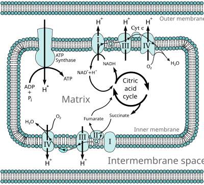
I mitokondrierne bliver vores mad omsat til energi i form af ATP. Dette sker via citronsyre cyklus og de fem mitokondrie komplekser. Basis viden om dette kan læses i wikipedia. Her ses citronsyre cyklus og de fem komplekser:


Figur fra Wikipedia: Oxidative phosphorylation
https://en.wikipedia.org/wiki/Oxidative_phosphorylation

I øverste venstre hjørne ses ATP synthase også kaldet complex V. Complex V sætter en fosfat-gruppe på ADP, så der dannes ATP. Complex V er yderligere beskrevet i Wikipedia: ATP synthase:  https://en.wikipedia.org/wiki/ATP_synthase

Professor Paul Fisher fra La Trobe Universitet har anvendt Seahorse teknologi til at undersøge mitokondrier fra ME patienter. Han har vist, at ved belastning arbejder complex V ineffektivt. Når cellerne ikke er belastede kan ATP produktionen følge med, men ved belastning går det galt. Effekten af complex V er nedsat med cirka 25% - mere hos de mest syge og mindre hos de mere raske ME patienter.

Complex V dysreguleringen medfører, at de andre mitokondrie komplekser arbejder mere for at prøve at kompensere. 

Professor Paul Fisher har fremlagt sine forskningsresultater i en præsentation:

Conclusions from Paul Fisher's "Specific Mitochondrial Respiratory Defects & Compensatory Changes in ME/CFS Patient Cells":
  • ATP synthesis by Complex V is less efficient in ME/CFS cells
  • Nonmitochondrial O2 consumption (metabolism) and mitochondrial respiratory capacity are elevated in ME/CFS cells
  • Respiration abnormalities correlate with disease state
  • Proteins involved in transporting molecules across the mitochondrial membrane and in mitochondrial fatty acid oxidation are upregulated in ME/CFS cells
  • TOR Complex I activity is elevated in ME/CFS lymphoblasts and could be responsible for upregulation of mitochondrial proteins
  • ME/CFS cells may be less able to respond to additional acute energy demands because they have already "turned up" their capacity as much as possible
  • Cause-effect relationships need to be determined


Her er link til præsentationen:
Specific Mitochondrial Respiratory Defects & Compensatory Changes in ME/CFS Patient Cells

Bloggen Health Rising har skrevet om præsentationen:
Emerging Insights #II: “The Cellular Equivalent of Chronic Fatigue” Found in ME/CFS

Artikel af Daniel Missailidis, Sarah Annesley og Paul Fisher
Pathological Mechanisms Underlying Myalgic Encephalomyelitis/Chronic Fatigue Syndrome
https://www.preprints.org/manuscript/201907.0196/v1

Hvis du vil vide mere om ATP synthase (complex V), så er her er review fra 2019:
ATP Synthase: Structure, Function and Inhibition

torsdag den 18. juli 2019

Betydning af acetyl-grupper for ME sygdomsmekanismen

ME patienter har dysreguleret glykolyse. McGregor et al (1) mener, at denne dysregulering medfører:

  1. nedsat dannelse af acetyl-grupper
  2. histone deacetylering
  3. nedsat acetylering af enzymer


McGregor et al artikel:
Post-Exertional Malaise Is Associated with Hypermetabolism, Hypoacetylation and Purine Metabolism Deregulation in ME/CFS Cases

McGregor holder foredrag om forskningen: An Omic Analysis of ME/CFS – an Assessment of Potential Mechanisms:
https://www.youtube.com/watch?v=UvGMbbyGtvg

Bloggen Health Rising har gennemgået forskningen:
Emerging Insights #1: McGregor’s Grand Conception of ME/CFS
https://www.healthrising.org/blog/2019/06/15/emerging-insights-mcgregor-metabolism-chronic-fatigue-glycolysis/


Nu følger lidt basisviden, om de begreber, der anvendes i artiklen/foredraget.

Acetyl-grupper

Acetat er saltet af eddikesyre. Den kemiske formel for acetat er  [CH3COO]−  og den kemiske formel for en acetyl-gruppe er   CH3CO,  Når en acetyl-gruppe sættes på en organisk forbindelse kaldes den en acetylering (engelsk: acetylation). Deacetylering er når acetyl-gruppen fjernes. Mange forskellige enzymer kan påsætte/fjerne acetyl-grupper i forskellige biokemiske sammenhænge.

Histoner

Histoner er de spoler, hvorpå vores DNA er oprullet. Tilgængeligheden af DNAet styres ved at påsætte/fjerne acetylgrupper.

HAT: Histone acetyltransferase sætter acetyl-grupper på histonerne
HDAC: Histone deacetylase fjerner acetyl-grupper fra histonerne

Læs om dette i wikipedia: Histone acetylation and deacetylation


Acetyl-CoA

Acetyl-CoA er et energi-rigt molekyle, som kan anvendes som brændstof i TCA-cyklus, men det anvendes også til at donere acetyl-grupper til histonerne. Acetyl-CoA dannes ved nedbrydning af pyruvat.

Her er link til en god figur, der viser anvendelsen af acetyl-CoA. Det er figur 3 fra reference 2.
https://www.ncbi.nlm.nih.gov/pmc/articles/PMC5934764/figure/embr201745432-fig-0003/

Det tyder på, at pyruvat dehydrogenase komplekset kan rykke ind i cellens kerne og producere acetyl-grupper til histonerne: Se figur 4 fra reference 2.
https://www.ncbi.nlm.nih.gov/pmc/articles/PMC5934764/figure/embr201745432-fig-0004/


Mere viden

Og hvis man vil forske i emnet, så er der fine metoder til det:
Integrated Analysis of Acetyl-CoA and Histone Modification via Mass Spectrometry to Investigate Metabolically Driven Acetylation

Ovenstående artikel har et let forståelig figur:


Referencer

1) Mcgregor et al: Post-Exertional Malaise Is Associated with Hypermetabolism, Hypoacetylation and Purine Metabolism Deregulation in ME/CFS Cases Diagnostics (Basel). 2019 Jul 4;9(3). pii: E70. doi: 10.3390/diagnostics9030070. https://www.ncbi.nlm.nih.gov/pubmed/31277442

2) Lisowski et al: Mitochondria and the dynamic control of stem cell homeostasis.
EMBO Rep. 2018 May;19(5). pii: e45432. doi: 10.15252/embr.201745432. Epub 2018 Apr 16.
https://www.ncbi.nlm.nih.gov/pubmed/29661859