Sider

fredag den 30. november 2018

Bile acid transporter SLCO3A1 and ME

Bile acid transporters maintain bile acid homeostasis.

Solute Carrier Organic Anion Transporter Family Member 3A1 (SLCO3A1) Is a Bile Acid Efflux Transporter in Cholestasis (1).

SLCO3A1 is up-regulated as an adaptive response to cholestasis (1).

Genome-wide association analysis identified a single nucleotide polymorphism (SNP) in SLCO3A1 in ME patients (2).

Epigenetic analysis identified that the gene SLCO3A1 (5'UTR) was hypomethylated in peripheral blood mononuclear cells (PBMC) from ME patients. (3).

Metabolomic analysis on plasma from ME patients identified lower levels of (4):

  • glycocholate
  • glycochenodeoxycholate
  • glycolithocholate
  • lithocholate
  • sulfoglycolithocholate
  • taurine

How is SLCO3A1 involved in ME?

Interestingly, bile acids activated receptors regulate innate immunity (5).

References

1) Pan et al. Solute Carrier Organic Anion Transporter Family Member 3A1 Is a Bile Acid Efflux Transporter in Cholestasis. Gastroenterology. 2018 Nov;155(5):1578-1592.e16. doi: 10.1053/j.gastro.2018.07.031. Epub 2018 Jul 29. https://www.ncbi.nlm.nih.gov/pubmed/30063921

2) Schlauch et al: Genome-wide association analysis identifies genetic variations in subjects with ME/CFS. 2016.doi.10.1038/tp.2015.208

3) Trivedi et al: Identification of ME/CFS - associated DNA methylation patterns.
Plos One 2018, 13, 7 https://journals.plos.org/plosone/article?id=10.1371/journal.pone.0201066

4) Germain et al: Metabolic profiling of a ME/CFS discovery cohort reveals disturbances in fatty acid and lipid metabolism. Mol. BioSyst. 2017, 13, 371 https://pubs.rsc.org/en/Content/ArticleLanding/2017/MB/C6MB00600K#!divAbstract

5) Fiorucci et al. Bile Acids Activated Receptors Regulate Innate Immunity. Front Immunol. 2018 Aug 13;9:1853. doi: 10.3389/fimmu.2018.01853. eCollection 2018.
https://www.ncbi.nlm.nih.gov/pubmed/30150987

onsdag den 28. november 2018

Lipid Transfer Proteins in ME

Lipid metabolism is dysregulated in ME patients (1, 2, 3).

Genes encoding lipid transfer proteins (LTPs) are epigentic changed in peripheral blood mononuclear cells (PBMC) from ME patients (4, 5, 6, 7).

Protein levels of LTPs are changed in the spinal fluid from ME patients (8).

Lipid Transfer Proteins

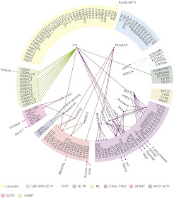
Within the eukaryotic cell, more than 1000 species of lipids define a series of membranes essential for cell function. Tightly controlled systems of lipid transport underlie the proper spatiotemporal distribution of membrane lipids, the coordination of spatially separated lipidmetabolic pathways, and lipid signaling mediated by soluble proteins that may be localized some distance away from membranes. Alongside the well-established vesicular transport of lipids, non-vesicular transport mediated by a group of proteins referred to as lipid-transfer proteins(LTPs) is emerging as a key mechanism of lipid transport in a broad range of biological processes. More than a hundred LTPs exist in humans and these can be divided into at least ten protein families. LTPs are widely distributed in tissues, organelles and membrane contact sites (MCSs), as well as in the extracellular space. They all possess a soluble and globular domain that encapsulates a lipid monomer and they specifically bind and transport a wide range of lipids (9).


Fig. 5. Lipid-transfer proteins have multiple modes of action (Figure from Chiappariono et al. ref 9)
a, LTPs can transfer lipids between cellular membranes and act as transporters. b, Some LTPs (chaperones) present lipids to an acceptor protein (e.g. enzymes, LTPs, transmembrane (TM) transporters or transcription factors). c, The LTD can be engaged in intramolecular interactions with other domains (illustrated here in purple) or proteins (not illustrated). Binding to the lipid cargo acts as a trigger that induces conformational changes and leads to the activation of signaling. This mechanism is sometimes coupled to the mechanisms described in a and b. LTPs have pleiotropic functions and can modulate lipid homeostasis, signaling and the structural organization of membranes.


Fig. 4. Domain organizations of human lipid-transfer proteins. The members of the ten families of LTPs are displayed.  (Figure from Chiapparino et al. ref 9).

PITPNA, PITPNC1 and PITPNM2 in ME

The genes encoding the phosphatidylinositol transfer proteins PITPNA, PITPNC1 and PITPNM2 were hypomethylated in PBMC from ME patients in Trivedi's study (7). PITPNM2 was hypomethylated in the genic regions: 1stExon, TSS200, TSS1500 and 3'UTR; and also showed changed DNA methylation pattern in 3 other studies (4, 5, 6).

GM2A in ME

A number of LTPs can transfer lipids to downstream enzymes. GM2 ganglioside activator (GM2A) is a lysomal LTP that works as a cofactor for the glycosphingolipid-degrading enzyme beta-hexosaminidase A (HEXA). GM2A is also involved in presentation of antigenic lipids by CD1 to T cells.

The GM2A gene promoter was hypomethylated in ME patients (table S7 in ref 7).

GM2A precursor, number of unique peptides identified in cerebrospinal fluid (table S1 in ref 8):
1) Controls: 12
2) ME patients: 32
3) Post  treatment  Lyme patients:32

HEXA chain precursor, number of unique peptides identified in cerebrospinal fluid (table S1 in ref 8):
1) Controls: 16
2) ME patients: 30
3) Post  treatment  Lyme patients:28


PLTP in ME

The gene encoding phospholipid transfer protein (PLTP) was hypomethylated (TSS1500) in PBMC from ME patients (7).

PLTP (TSS1500) was differentially methylated in PBMC from ME patients subtypes (6).

PLTP precursor, number of unique peptides identified in cerebrospinal fluid (table S1 in ref 8):
1) Controls: 16
2) ME patients: 31
3) Post  treatment  Lyme patients: 26

STARD13 and ME

The gene encoding StAR related lipid transfer domain containing 13 (STARD13) was hypermethylated in genic region 3'UTR and hypomethylated in TSS200 in PBMC from ME patients (7).

STARD13 was differentially methylated in PBMC from ME patients subtypes (6).

This was some of the LTPs involved in ME.

References

1) Naviaux RK, Naviaux JC, Li K, Bright AT, Alaynick WA, Wang L, Baxter A, Nathan N et al (2016) Metabolic features of chronic fatigue syndrome. Proc Natl Acad Sci U S A 113:E5472–E5480. https://doi.org/10.1073/pnas.1607571113 

2) Germain et al: Metabolic profiling of a ME/CFS discovery cohort reveals disturbances in fatty acid and lipid metabolism. Mol. BioSyst. 2017, 13, 371 https://pubs.rsc.org/en/Content/ArticleLanding/2017/MB/C6MB00600K#!divAbstract

3) Nagy-Szakal et al: Insights into ME/CFS phenotypes through comprehensive metabolomics. Nat. Sci. Rep, 2018, 8. https://www.nature.com/articles/s41598-018-28477-9

4) de Vega et al: DNA methylation Modifications associated with CFS. PlosOne, 2014, 9, 8. https://journals.plos.org/plosone/article?id=10.1371/journal.pone.0104757 

5) de Vega et al: Epigenetic modifications and glucocorticoid sensitivity in ME/CFS. BMC Medical Genomics, 2017, 10, 11 https://www.ncbi.nlm.nih.gov/pmc/articles/PMC5324230/

6) de Vega et al: Integration of DNA methylation & health scores identifies subtypes in ME/CFS. Epigenomics 2018, 10, 5 https://www.futuremedicine.com/doi/full/10.2217/epi-2017-015

7) Trivedi et al: Identification of ME/CFS - associated DNA methylation patterns.
Plos One 2018, 13, 7 https://journals.plos.org/plosone/article?id=10.1371/journal.pone.0201066

8) Schutzer et al: Distinct Cerebrospinal Fluid Proteomes Differentiate Post- Treatment Lyme Disease from Chronic Fatigue Syndrome. PLOS One February 2011, volume 6, Issue  https://journals.plos.org/plosone/article?id=10.1371/journal.pone.0017287

9) Chiapparino et al: The orchestra of lipid-transfer proteins at the crossroads between metabolism and signaling. Prog Lipid Res. 2016 Jan;61:30-9. doi: 10.1016/j.plipres.2015.10.004 https://www.ncbi.nlm.nih.gov/pubmed/26658141

fredag den 23. november 2018

Red blood cell deformability, metabolism and extracellular vesicles in ME/CFS

Red blood cell deformability

Red blood cells (RBCs; erythrocytes) are typically biconcave in shape, transport hemoglobin-bound oxygen and are reversibly deformable facilitating trafficking through capillaries. Decreased deformability of RBCs adversely affects tissue oxygenation (1).

RBC deformability is reduced in some ME/CFS patients (2).

Metabolism and red blood cell membrane

The cell membranes in red blood cells are made up of proteins, fats, and carbohydrates. This means that abnormalities in the cell membrane can be a good way to spot disordered metabolism of these nutrients. Issues with producing too much of a certain metabolite, or not being able to break down a nutrient can cause abnormalities in the cell membrane, which lead to irregular shapes of RBCs (2).

Erythrocyte omega-3 index (5.75%) and n-3 PUFA levels are low in individuals with CFS/ME (3).

Extracellular vesicles in sepsis decrease RBC deformability

A growing body of evidence suggests that extracellular vesicles (EVs) play a role in cell-to-cell communication, and are involved in both physiological and pathological processes (4).

A study on mice showed a significant decrease in RBC deformability following sepsis. Extracellular vesicles isolated from the plasma of mice with sepsis significantly decreased deformability of RBCs ex vivo (1).

Extracellular vesicles in blood from ME patients have been analyzed: The amount of EV-enriched fraction was significantly higher in CFS/ME subjects than in healthy controls (HCs) (p = 0.007) and that EVs were significantly smaller in CFS/ME patients (p = 0.014). Circulating EVs could be an emerging tool for biomedical research in CFS/ME. These findings provide preliminary evidence that blood-derived EVs may distinguish CFS/ME patients from HCs (4).

I suggest a pilot study:

ME/CFS vesicles put together with red blood cells from healthy controls. Can microvesicles from ME/CFS patients decrease deformability of RBCs from healthy controls in vitro?


References


1) Subramini et al. Effect of plasma-derived extracellular vesicles on erythrocyte deformability in polymicrobial sepsis. Int Immunopharmacol. 2018 Oct 16;65:244-247. doi: 10.1016/j.intimp.2018.10.011. https://www.ncbi.nlm.nih.gov/pubmed/?term=30340103

2) Erythrocyte Deformability As a Potential Biomarker for Chronic Fatigue Syndrome. Amit K Saha, Brendan R Schmidt, Julie Wilhelmy, Vy Nguyen, Justin Do, Vineeth C Suja, Mohsen Nemat-Gorgani, Anand K Ramasubramanian and Ronald W Davis

Blood 2018 132:4874; doi: https://doi.org/10.1182/blood-2018-99-117260 http://www.bloodjournal.org/content/132/Suppl_1/4874

OMF-funded research: red blood cell deformability in ME/CFS
https://www.omf.ngo/2018/03/21/omf-funded-research-red-blood-cell-deformability-in-me-cfs/

RBC shape, RBC deformability
https://www.omf.ngo/2018/04/04/rbc-shape-rbc-deformability/

3) Castro-Marrero et al: Low omega-3 index and polyunsaturated fatty acid status in patients with chronic fatigue syndrome/myalgic encephalomyelitis.
Prostaglandins, Leukotrienes and Essential Fatty Acids
Volume 139, December 2018, Pages 20-24
https://www.sciencedirect.com/science/article/pii/S095232781830053X

4) Castro-Marrero et al:
Circulating extracellular vesicles as potential biomarkers in chronic fatigue syndrome/myalgic encephalomyelitis: an exploratory pilot study. J Extracell Vesicles. 2018 Mar 22;7(1):1453730. doi: 10.1080/20013078.2018.1453730. eCollection 2018. https://www.ncbi.nlm.nih.gov/pubmed/29696075

tirsdag den 6. november 2018

NPY, AgRP, POMC and NUCB2 in ME

Hypothalamic Pro-opiomelanocortin (POMC) and Neuropeptide Y/Agouti-Related Peptide (NPY/AgRP) neurons are critical nodes of a circuit within the brain that sense key metabolic cues as well as regulate metabolism. Importantly, these neurons retain an innate ability to rapidly reorganize synaptic inputs and electrophysiological properties in response to metabolic state (1).

Exercise (single bout and/or chronic training) increases insulin sensitivity leading to improved insulin stimulated glucose uptake in muscle and reduced basal hepatic glucose production . Within the arcuate nucleus, the melanocortin system is an interface between signals of metabolic state and neural pathways governing energy balance and glucose metabolism. In particular, the orexigenic neuropeptide Y/Agoutirelated peptide (NPY/AgRP) neurons are activated in response to food deprivation, while the anorexigenic proopiomelanocortin (POMC)- expressing cells are inhibited . In addition to contributing to energy balance, the activity of arcuate NPY/AgRP and POMC neurons also have profound effects on glucose metabolism. These changes in cellular activity have been attributed to both native channel properties as well as (re)organization of synaptic connectivity (1 and references herein).

New research shows: Cellular and synaptic reorganization of arcuate NPY/AgRP and POMC neurons after exercise (1).

Nesfatin-1 is an 82–amino acid polypeptide derived from the precursor protein nucleobindin 2 (NUCB2), whose processing also yields nesfatin-2 and -3, two peptides with so far unknown functions (2).

Nesfatin-1 is involved in several processes including modulation of gastrointestinal functions, energy metabolism, glucose and lipid metabolism, thermogenesis, mediation of anxiety and depression, as well as cardiovascular and reproductive functions (2).

Use the link to se figure with NUCB2 and the hypothalamus (3): 

As you can se in the figure, the hypothalamus is a key brain area for maintaining glucose and energy homeostasis via the ability of hypothalamic neurons to sense, integrate, and respond to numerous metabolic signals.

Mitochondrial function has emerged as an important component in the regulation of hypothalamic neurons controlling glucose and energy homeostasis. Although the underlying mechanisms are not fully understood, emerging evidence indicates that mitochondrial dysfunction in hypothalamic neurons may contribute to the development of various metabolic diseases (4).

NPY, AgRP, POMC and NUCB2 in ME

NPY precursor, number of unique peptides identified in cerebrospinal fluid (table S1 in ref 5):
1) Controls: 4
2) ME patients: 7
3) Post  treatment  Lyme patients: 6

The plasma level of neuropeptide Y is elevated in ME patiens (6).

POMC, number of unique peptides identified in cerebrospinal fluid (table S1 in ref 5):
1) Controls: 1
2) ME patients: 2 
3) Post  treatment Lyme patients: 1

Some ME patients have single nucleotide plymorphism (SNP) in POMC (7).

The gene POMC (TSS1500, 5'UTR) is hypomethylated in peripheral blood mononuclear cells  (PBMC) from ME patients (8).

NUCB1 precursor, number of unique peptides identified in cerebrospinal fluid (table S1 in ref 5):
1) Controls: 29
2) ME patients: 43
3) Post  treatment Lyme patients: 51

NUCB2 precursor, number of unique peptides identified in cerebrospinal fluid (table S1 in ref 5):
1) Controls: 9
2) ME patients: 8
3) Post  treatment Lyme patients: 13

The gene NUCB2 (5'UTR) is hypomethylated in PBMC from ME patients (8).

The gene NUCB2 (5'UTR) is differentially methylated in PBMC from ME patient subtyes (9).


References

1) He et al: Cellular and synaptic reorganization of arcuate NPY/AgRP and POMC neurons after exercise. Mol Metab. 2018 Sep 12. pii: S2212-8778(18)30870-6. doi: 10.1016/j.molmet.2018.08.011 https://www.ncbi.nlm.nih.gov/pubmed/30292523

2) Schalla and Stengel: Current Understanding of the Role of Nesfatin-1. J Endocr Soc. 2018 Oct 1; 2(10): 1188–1206. Published online 2018 Sep 10. doi: [10.1210/js.2018-00246]
https://www.ncbi.nlm.nih.gov/pmc/articles/PMC6169466/

3) Figure from:  Diabetes. 2012 Aug; 61(8): 1920–1922.
and https://www.ncbi.nlm.nih.gov/pubmed/22826310

4) Jin and Diano: Mitochondrial Dynamics and Hypothalamic Regulation of Metabolism. Endocrinology, Volume 159, Issue 10, 1 October 2018, Pages 3596–3604,https://doi.org/10.1210/en.2018-00667 https://academic.oup.com/endo/article-abstract/159/10/3596/5091402?redirectedFrom=fulltext

5) Schutzer et al: Distinct Cerebrospinal Fluid Proteomes Differentiate Post- Treatment Lyme Disease from Chronic Fatigue Syndrome. PLOS One February 2011, volume 6, Issue  https://journals.plos.org/plosone/article?id=10.1371/journal.pone.0017287

6) Fletcher et al: Plasma neuropeptide Y: a biomarker for symptom severity in chronic fatigue syndrome. Behav Brain Funct. 2010 Dec 29;6:76. doi: 10.1186/1744-9081-6-76 https://www.ncbi.nlm.nih.gov/pubmed/21190576

7) Smith et al: Polymorphisms in genes regulating the HPA axis associated with empirically delineated classes of unexplained chronic fatigue. PHARMACOGENOMICSVOL. 7, NO. 3COLLABORATIVE STUDY: CHRONIC FATIGUE SYNDROME – RESEARCH REPORT
https://www.futuremedicine.com/doi/10.2217/14622416.7.3.387

8) Trivedi et al: Identification of ME/CFS - associated DNA methylation patterns.
Plos One 2018, 13, 7 https://journals.plos.org/plosone/article?id=10.1371/journal.pone.0201066

9) de Vega et al: Integration of DNA methylation & health scores identifies subtypes in ME/CFS. Epigenomics 2018, 10, 5 https://www.futuremedicine.com/doi/full/10.2217/epi-2017-015